Accurate Diagnosis of TMJ Pain and Dysfunction (Preview)

Disclaimer: Please read the Disclaimer at the bottom of this page.
Copyright © Educom Pty Ltd: All content on the educomce.com website, including text, graphics, videos, and downloadable files, is the property of Educom Continuing Education, a division of Educom Pty Ltd, and is protected by copyright and other intellectual property laws under international conventions. Unauthorized use or duplication of this material without express written permission from Educom Pty Ltd is strictly prohibited.
Content Links
The following buttons can be used to navigate the content on this web page.
Diagnostic Features
Temporomandibular Joint Pain and Dysfunction
Introduction
Temporomandibular Joint Disorders
Temporomandibular joint disorders are commonly seen in clinical practice. They are reported to be more common in women and have a peak age incidence of 20–40 years. Clinical presentations of temporomandibular joint disorders are characterized by acute or chronic pain and may include the following:
-
- Temporomandibular joint dysfunction
- Facial pain
- Ear pain, a feeling of ear fullness, and tinnitus
- Neck pain
- Orbital pain
- Dizziness
Temporomandibular Joint Dysfunction
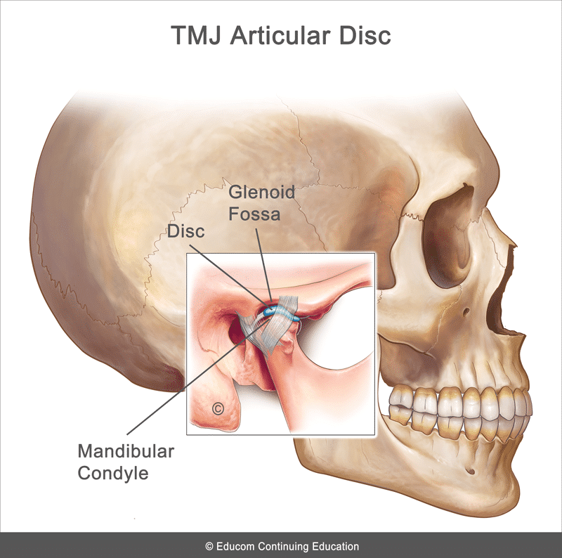
Temporomandibular joint dysfunction is characterized by pain arising from an abnormal relationship between the temporomandibular joint (TMJ) articular disc and the adjacent articular surfaces, as well as the surrounding myofascial structures. The TMJ is an extremely important articulation necessary for mastication, swallowing, facial expression, and communication. This joint is classified as a ginglymoarthrodial joint allowing essentially a hinge-like movement (rotation) combined with a gliding motion (translation). On opening the mouth, the movement begins with rotation of the mandibular condyle in the glenoid fossa, followed by a forward translation of the condyles. The TMJ involves articulation between the mandibular condyle and the glenoid fossa of the temporal bone. These two osseous structures are separated by a fibrocartilagenous disc. The TMJ is strengthened by its joint capsule and several ligaments (the sphenomandibular, stylomandibular, pterygomandibular, malleolomandibular and collateral ligaments).
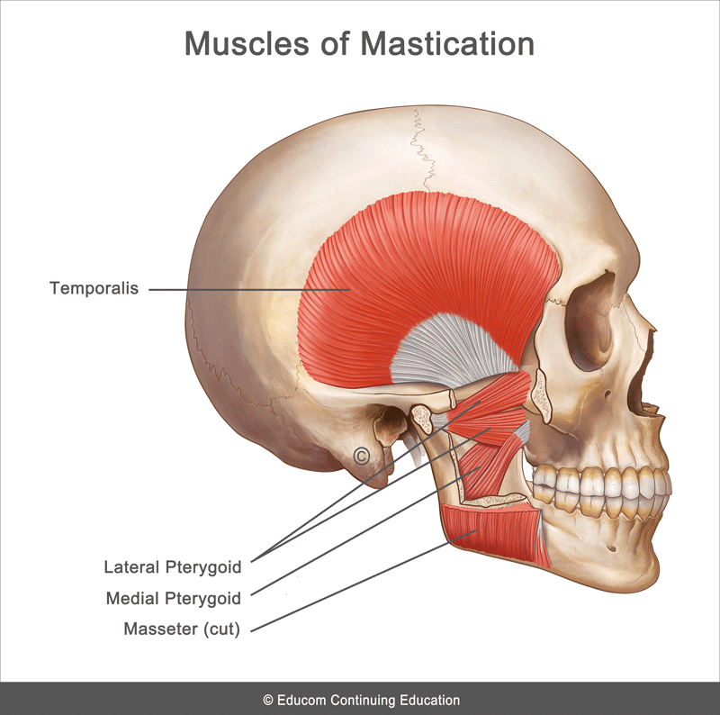
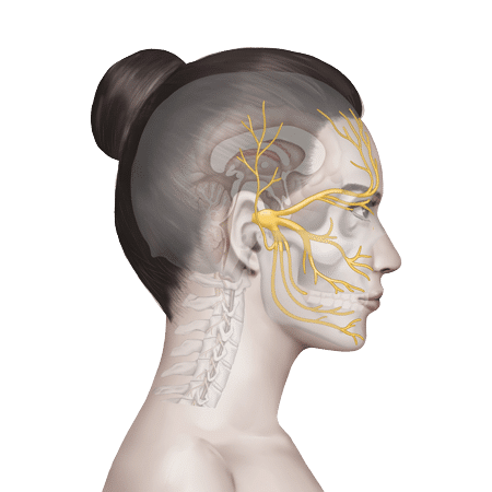
The muscles involved in the opening and closing of the mouth are the primary muscles of mastication, including the masseter, temporalis, and medial pterygoid muscles which elevate the mandible to close the mouth, and the lateral pterygoid muscle which assists in opening the mouth by guiding forward movement of the jaw. Innervation of the TMJ and its associated muscles involves branches of the third division of the trigeminal nerve.
The etiology of TMJ dysfunction is thought to be multifactorial including anatomical, pathophysiological, and psychosocial factors. Musculoskeletal dysfunction is the most common contributing factor to TMJ dysfunction. This could be due to the following:
-
- Joint trauma
- Muscle imbalance
- Poor head and neck posture
- Myofascial pain syndromes
- Internal joint derangement
- Degenerative joint disease
- Chronic bruxism (clenching or grinding the teeth)
- Dental malocclusion
- A physical manifestation of a psychological disorder (e.g., depression, anxiety, or post-traumatic stress disorder)
Differential Diagnosis of Orofacial Pain
Patients presenting with orofacial pain may be suffering from several conditions. The differential diagnosis includes the following:
-
- TMJ dysfunction
- Dental caries, abscess, or malocclusion
- Otitis media and otitis externa
- Upper cervical facet joint dysfunction
- Mastoiditis
- Migraine headache
- Cluster headache
- Tension-type headache
- Trigeminal neuralagia
- Post-herpetic neuralgia
- Giant cell arteritis (Temporal arteritis)
- Parotitis
- Mandibular fracture or dislocation
- Sinusitis
- Cancer of the jaw, head, or neck
History
- Preauricular pain that is usually described as deep and aching with sharp exacerbations on jaw movement
- Pain that may refer to the head and neck
- Pain that is aggravated by chewing, yawning, or talking for extended periods
- Joint clicking, popping, or snapping on jaw movement
- Limited jaw opening
- May have jaw-locking
- Headache
- May have associated otological symptoms (e.g., tinnitus, vertigo, earache, or hearing loss)
Physical Examination
- Abnormal mandibular movement
- Decreased TMJ range of motion (inability to fully open the mouth, typically 25 mm or less)
- Local tenderness over the joint and/or in the muscles of mastication
- Palpation may reveal a clicking or popping sensation with jaw movement
- Palpation may reveal grinding or crepitus with jaw movement
- Muscle tension or spasm
- Pain on jaw clenching due to dynamic loading
- May have evidence of tooth wear or malocclusion
- May have abnormal cervical posture
Diagnostic Imaging
The diagnosis of TMJ dysfunction is usually clinical. Plain radiography can help to rule out degenerative joint disease, fractures, dislocations, and bone pathology. Magnetic resonance imaging (MRI) is considered the gold standard for the investigation of TMJ disorders as it is able to assess soft tissue structures, articular disc displacement, and the presence of joint effusion.
Red Flags
The following are examples of red flags for patients presenting with orofacial pain:
-
- A history of significant injury
- Severe pain
- Unrelenting pain
- Nocturnal pain
- Unexplained weight loss
- Fever
- Deformity
- Trismus
- Significant swelling
- Unilateral hearing loss or a new onset of tinnitus
- Vestibular dysfunction
- Significant loss of range of motion
- Severe tenderness on palpation or severe pain with any examination procedure
If any red flags are identified during history taking and clinical examination, referral for urgent medical evaluation and further investigation is warranted.

TMJ Palpation
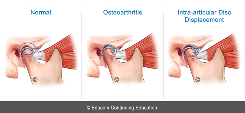
When palpating the TMJ (e.g., by placing fingers in the patient’s ears while they open and close their mouth), if a crackling or grating sound or sensation is present, this suggests osteoarthritis. If a clicking sensation is present, this could indicate displacement of the intra-articular disc.
Innervation of the TMJ and Associated Muscles
Since innervation of the TMJ and its associated muscles involves branches of the third division of the trigeminal nerve, pain from mandibular pathology or the mandibular teeth can be referred to the preauricular area and misinterpreted as arising from TMJ dysfunction. On the other hand, pain arising from the TMJ may be perceived by the patient as an earache.
Myofascial Pain Syndrome Presenting as TMJ Pain
In patients presenting with TMJ pain, a complete clinical examination should include an evaluation for the presence of myofascial trigger points as either the primary cause of the pain or as a concomitant condition. The primary muscles to evaluate include the following:
-
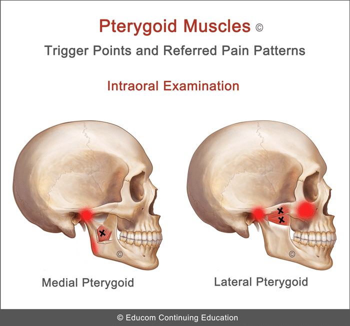
- Medial Pterygoid: Pain primarily in the region of TMJ
- Lateral Pterygoid: Pain primarily in the region of TMJ and maxillary area sometimes resembling as “sinus pain”
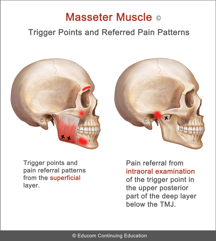
- Masseter – Superficial Layer: Pain primarily in the lowewr jaw and zygomatic arch
- Masseter – Deep Layer: Pain primarily in the TMJ (trigger point palpated intraorally)
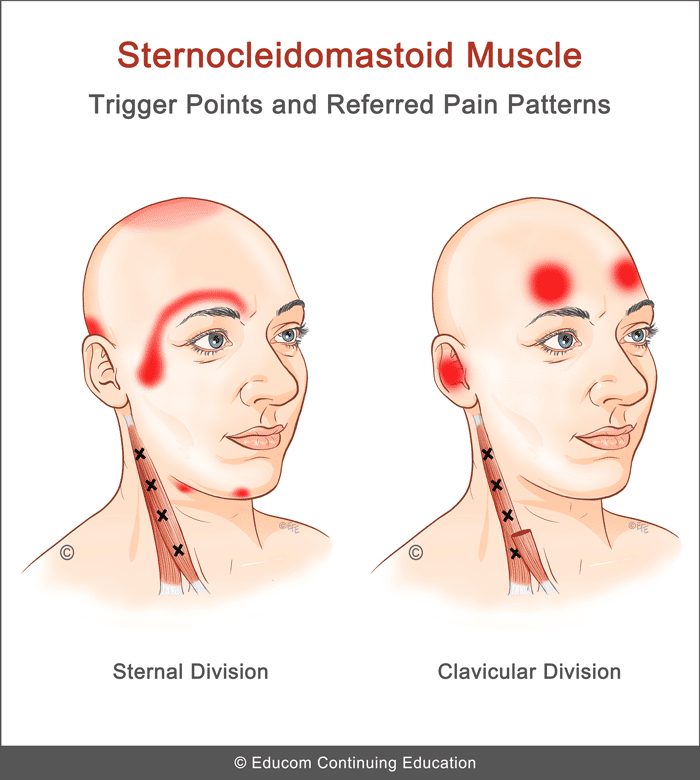
- Sternocleidomastoid – Clavicular Division: Pain primarily in the forehead (may refer to both sides), pain deep in the “ear” with/without feeling of lightheadedness and imbalance
- Sternocleidomastoid – Sternal Division: Pain primarily in the temporal region and deep behind the eye
Management
The suggested conservative therapy, home advice, and clinical tips in this section are based on published materials and the clinical experience of the authors of this course. This should not be interpreted as a prescriptive guide to the treatment of this or any other condition. The use of this content is subject to the “Disclaimer” found at the bottom of this web page.

Acute Phase
-
- Evaluation and Correction of Joint Dysfunction (if present) – If indicated, any management techniques that are applied should minimize excessive loading.
- Low Level Laser Therapy – Some patients benefit from the application of low level laser therapy over the involved TMJ to assist with pain reduction and healing.
- Myofascial Release – If any myofascial pain syndrome is present, treat the trigger points using myofascial release, particularly in the head and neck regions.
- Acupuncture – Some patients benefit from acupuncture treatment.
- Ice – Application of ice over the involved TMJ. The ice pack may be applied for 5 to 10 minutes as often as every hour.
Subacute and Rehabilitation Phase
-
- Correction of Joint Dysfunction (if present)
- Low Level Laser Therapy
- Acupuncture
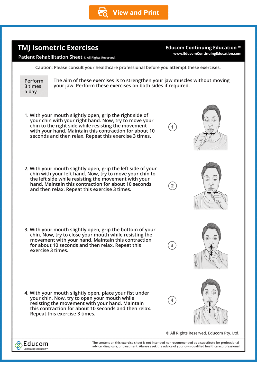
- TMJ Isometric Exercises
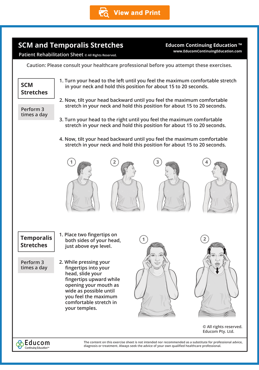
- SCM and Temporalis Stretches
- Ice – Application of ice if the pain returns.

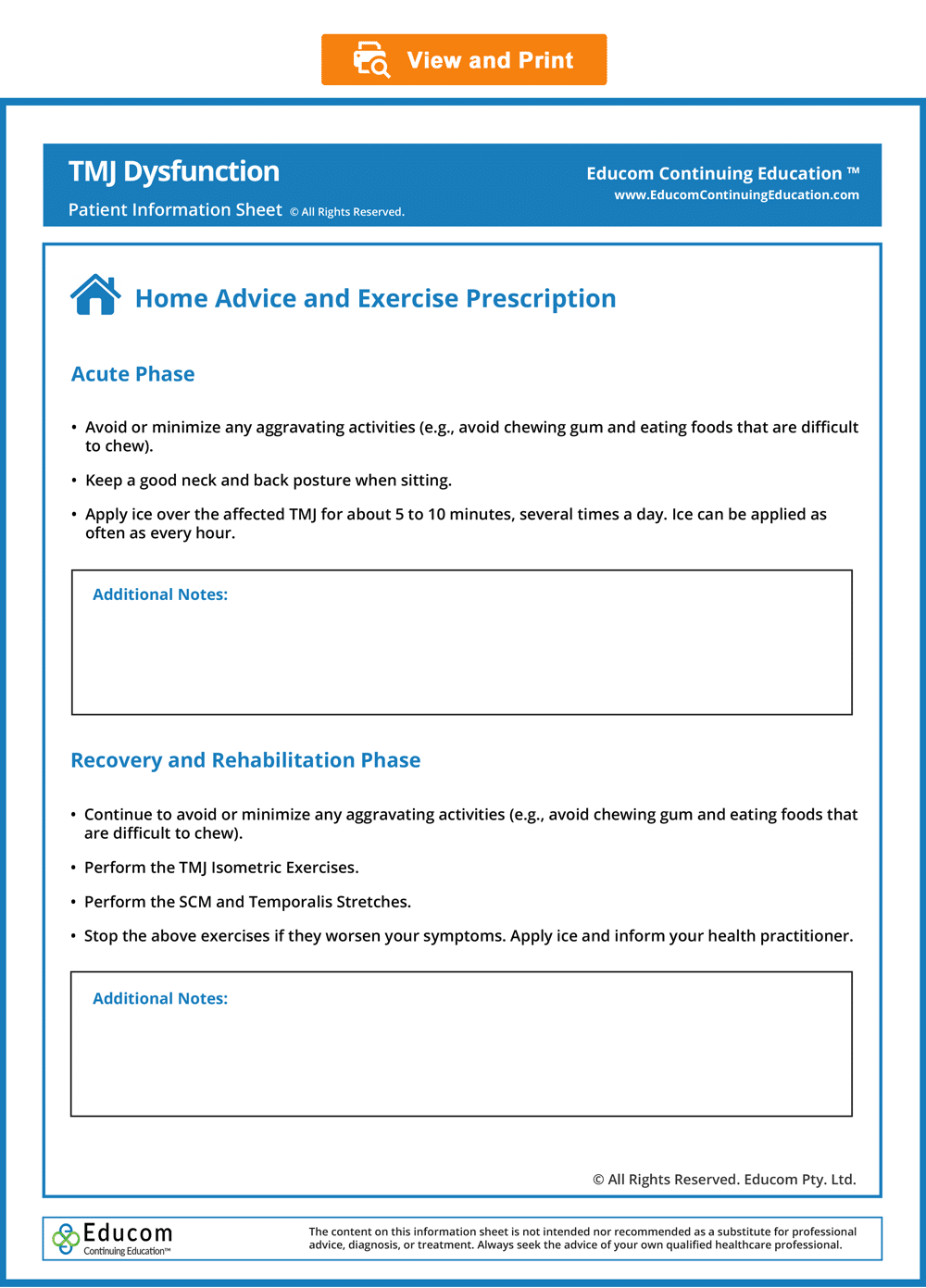
Acute Phase
-
- Avoid or minimize any aggravating activities (e.g., avoid eating foods that are difficult to chew, including chewing gum).
- Keep a good neck and back posture when sitting.
- Apply ice over the affected TMJ for about 5 to 10 minutes, several times a day. Ice can be applied as often as every hour.
Subacute and Rehabilitation Phase
-
- Continue to avoid or minimize any aggravating activities (e.g., avoid eating foods that are difficult to chew, including chewing gum).
- Perform the TMJ Isometric Exercises.
- Perform the SCM and Temporalis Stretches
- Stop these exercises if they worsen your symptoms and report it to your practitioner.
- Apply ice if your TMJ pain returns or is aggravated.
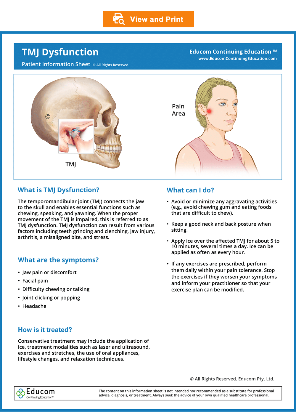
For Patients
Patient Information, Home Advice, and Exercise Prescription Sheets
- You may wish to give your patients the following downloadable and ready-to-use Patient Information, Home Advice and Exercise Prescription sheets. In our experience, patients who are well informed about their condition and the recommended management are more likely to achieve good outcomes, become loyal patients, and recommend their family and friends to seek treatment.
- To view, download, or print these sheets, simply click on the images below.
© Educom Pty. Ltd. All Rights Reserved.
Patient Exercise Sheets
- When appropriate, the patient should gradually begin doing their prescribed exercises at home. Always recommend warm-up activities before commencing specific exercises. Warm-up activities include simple limbering movements or prescribed strength exercises at light loads.
- If stretches are indicated, instruct your patients to perform them after they have completed their strengthening exercises.
- Always recommend warm-up activities before commencing specific exercises. Warm-up activities include simple limbering movements or prescribed strength exercises at light loads.
- Always instruct your patients to use caution when performing their exercises in order to avoid overloading, overstretching, or any undue pain.
- To view, download or print the Patient Exercise Sheet, simply click on the image below.
© Educom Pty. Ltd. All Rights Reserved.


To encourage critical thinking and help you sharpen your diagnostic reasoning skills, we recommend the following steps:
Step 1: Read through the provided information for each section of the case scenario.
Step 2: Consider the “Diagnostic Reasoning Question” for that section and attempt to answer the question before reading our suggested answer. When ready, click “View Suggested Answer” to compare your answer with ours.
Please note that the suggested answers are only indicative and not exhaustive. They serve as guidelines and should not be considered complete answers.
Patient’s History
In the sections below, you are provided with case history information. The material presents a systematic approach to performing a focused and relevant case history in order to narrow down the possible causes of the patient’s complaint. As you read the following material, you are encouraged to answer the questions posed to test your diagnostic reasoning skills.
Who is the patient and where is the pain?
When was the onset and what caused the onset?
Your patient is a 32-year-old female teacher who presents with pain in the right side of her face, which has been present for about two weeks. She points to the area in front of her right ear as the primary site of her pain. She first became aware of the pain within 24 hours following a dental consultation that required a prolonged procedure to repair a cracked tooth. She has since returned to the dentist, who recommended taking painkillers and giving it time to resolve.
|
What differential diagnoses would you consider for this patient? |
View Suggested Answer
Pain in the preauricular region can indicate several conditions, including:
-
- Temporomandibular joint dysfunction
- Myofascial pain syndromes (e.g., the medial and lateral pterygoids, masseter, and sternocleidomastoid muscles)
- Upper cervical facet joint dysfunction
- Dental caries, abscess, or malocclusion
- Otitis media and otitis external
- Trigeminal neuralgia
- Post-herpetic neuralgia
- Giant cell arteritis (temporal arteritis)
- Parotitis
- Mandibular fracture or dislocation
- Cancer of the jaw, head, or neck
|
Based on the patient’s history, how would you differentiate between trigeminal neuralgia and temporomandibular joint (TMJ) dysfunction? |
View Suggested Answer
-
- Trigeminal Neuralgia – In trigeminal neuralgia, the patient typically describes the pain as sudden, severe, stabbing, electric shock-like pain lasting from a few seconds to a few minutes. Pain is usually localized to specific branches of the trigeminal nerve, often affecting one side of the face. Light touch or movement may trigger pain.
-
- TMJ Dysfunction – In TMJ Dysfunction, pain is often localized to the temporomandibular joint but may radiate to surrounding areas, including the jaw, temples, or even the neck. Pain may be exacerbated by chewing, clenching the jaw, or other jaw movements. Patients may report clicking or popping sounds in the jaw joint, limited jaw movement, muscle stiffness or tenderness in the jaw or face, and occasionally headaches.
What are the pain characteristics?
What are the aggravating and relieving factors?
What has been the course of the pain?
She describes the pain as a constant dull ache with sharp exacerbations brought on by jaw movement, particularly when chewing or yawning. She rates the dull ache as a 3 out of 10 and the sharp exacerbations as a 9 out of 10 in intensity. She has found that applying an ice pack directly over the area of pain gives her temporary relief. She took painkillers on the dentist’s advice, which also gave her temporary relief, but she has stopped taking them, preferring to get your assistance to resolve the problem instead. There has been no change in the intensity of her pain since the onset.
|
What are some potential causes or underlying conditions that could lead to a patient experiencing a constant dull ache in the jaw with sharp exacerbations triggered by jaw movement, specifically when chewing or yawning? |
View Suggested Answer
Her description of pain aggravated by jaw movement strongly suggests the involvement of the TMJ, the muscles of mastication, or a dental problem. However, it may also indicate the presence of an external or middle ear infection.
|
How does the temporary pain relief through ice packs and painkillers inform our understanding of the underlying condition? |
View Suggested Answer
The relief of pain through the application of an ice pack may suggest an inflammatory process, though it could also result from an analgesic effect similar to that provided by painkillers.
Are there any associated symptoms?
She says that the sharp exacerbations of pain are accompanied by a clicking sensation in her right jaw when she chews food or yawns. The dull ache at her jaw sometimes spreads to the right side of her head, particularly after a full day of teaching. In addition, she complains of a mild right earache and thinks that the hearing in her right ear may be slightly diminished.
|
How does clicking in the jaw accompanied by pain inform the diagnostic process? |
View Suggested Answer
An occasional jaw click that is not painful is common in the general population. However, dysfunction of the TMJ, including issues such as disc displacement or joint degeneration, can cause sharp exacerbations of pain and clicking sensations during jaw movements. Clicking sensations accompanied by pain when chewing may also be due to changes in bite alignment arising from dental issues.
|
How does the spread of jaw pain to the side of the head and the development of associated symptoms such as mild earache and diminished hearing inform the diagnostic process? |
View Suggested Answer
A dull ache at the jaw that spreads to the side of the head, particularly after prolonged jaw movement (e.g., a day of talking), is consistent with referred pain from TMJ dysfunction. Dysfunction in the TMJ can also lead to pain that refers to adjacent areas, including the head and neck. However, myofascial pain syndrome, dental problems, and ear infections could also cause these referral patterns. Perceived hearing loss may indicate an ear infection, but this finding has also been reported in TMJ dysfunction.
Is there a past history that is relevant to the current complaint?
She has no past history of jaw pain.
|
How does the absence of a previous history of jaw pain inform the diagnostic process? |
View Suggested Answer
Her lack of a previous history of jaw pain suggests that the current issue may be acute or caused by a recent event, rather than being a chronic or recurring problem.
Are there any “red flags”?
The patient is asked the following questions to identify any “red flags” that could indicate serious pathology. Even if the patient has already provided information in the case history that relates to these questions, it is recommended that they be readdressed to ensure a thorough exploration.
-
- Do your symptoms disappear even for a short time? “Yes. I am not aware of the pain when I first wake up in the morning.”
- Does the pain wake you up at night? “No.”
- Are you experiencing any dizziness or loss of balance? “No.”
- Have you recently had any fever, chills, or night sweats? “No.”
- Have you recently had an infection or other illness? “No.”
- Have you had any unexplained weight loss recently? “No.”
- Do you have a history of cancer, inflammatory arthritis, or fracture arising from minor trauma? “No.”
|
Do any of the patient’s answers raise a red flag? |
View Suggested Answer
None of the patient’s responses raise a red flag.
What is the list of possible causes for the patient’s complaint?
|
How would you rank the differential diagnosis list based on the available history? |
View Suggested Answer
-
- Temporomandibular joint dysfunction
- Myofascial pain syndromes (e.g., the medial and lateral pterygoids, masseter, and sternocleidomastoid muscles)
- Dental malocclusion
- Otitis media and otitis external
- Upper cervical facet joint dysfunction
Reflection Point
Please stop and take a moment to consider whether the main requirements of an adequate and relevant patient history taking have been fulfilled. Are there any additional questions you would have asked and, if so, why?
Before the physical examination findings are presented below, please reflect on what physical examination procedures you would perform to adequately evaluate this patient.
Performing Physical Examination
In the sections below, you are provided with physical examination findings for this patient. The material presents a systematic approach to performing a focused and relevant physical examination to narrow down the possible causes of the patient’s complaint. You are encouraged to answer the questions posed to test your clinical reasoning and diagnostic skills as you read the following material. As you read the following material, you are encouraged to identify whether the essential elements of the physical examination have been adequately covered.
Vital Signs
Her vital signs are within normal limits.
|
What do normal vital signs indicate? |
View Suggested Answer
Normal vital signs typically indicate that there are no immediate life-threatening conditions. Normal temperature, in particular, indicates the absence of acute infection.
Inspection
Inspection of the right preauricular area reveals no evidence of swelling, redness, or deformity. Inspection of the right ear, including an otoscopic examination, is normal. Inspection of her head and neck posture is also normal.
|
How does the absence of visible swelling, redness, or deformity in the preauricular area inform the diagnostic process? |
View Suggested Answer
The absence of swelling, redness, or deformity in the preauricular area helps to rule out local inflammation, infection, or osseous pathology.
|
How do normal results from an ear inspection and otoscopic examination influence the diagnostic process? |
View Suggested Answer
Normal ear examination helps rule out otitis media and otitis externa.
Range of Motion
TMJ Active Movement Assessment reveals a reduction in jaw opening, measuring only 25 mm, with her jaw deviating to the right and resulting in right TMJ pain. When asked to move her jaw from side to side, she has limited movement to the left side, which also results in pain in the right TMJ. Assessment of the cervical active and passive ranges of motion is found to be normal.
|
What do the TMJ Active Movement Assessment findings indicate? |
View Suggested Answer
The findings indicate TMJ dysfunction caused by limited mandibular opening due to either:
-
- Hypertonicity or imbalance in the muscles of mastication
- Displacement of the TMJ meniscus resulting in interference with the normal forward movement of the mandibular condyle
- TMJ pathology (e.g., arthritis or local osseous pathology)
The absence of findings on assessment of the cervical ranges of motions helps to rule out cervical spine involvement.
Please watch the video below if you wish to see how the TMJ Active Movement Assessment is performed.
The Resisted Jaw Movement Assessment reveals an aggravation of the patient’s pain on resisted jaw closure and resisted jaw deviation to the left.
|
How do the Resisted Jaw Movement Assessment findings inform the diagnostic process? |
View Suggested Answer
These findings are further evidence of TMJ dysfunction either due to an intraarticular disorder or the involvement of the muscles of mastication.
Please watch the video below if you wish to see how the Resisted Jaw Movement Assessment is performed.
Palpation
The right TMJ is tender on palpation. While palpating, jaw opening and closing reveal a clicking sensation accompanied by pain. In addition, it is noted that the right mandibular condyle is not sliding forward adequately. Muscle palpation reveals tenderness in the right medial and lateral pterygoid, temporalis, and masseter muscles. No nodules or pain referral to the TMJ is found on examination of the masseter or the SCM muscles. The right ear, mastoid process, and parotid gland are non-tender on palpation. A functional examination of the cervical vertebral motion segments is normal.
|
How do palpation findings of tenderness in the right TMJ, pain accompanied by a clicking sensation during jaw movement, and insufficient forward sliding of the right mandibular condyle collectively contribute to the diagnostic process? |
View Suggested Answer
The TMJ tenderness, the clicking sensation, and the inadequate forward movement of the right mandibular condyle provide strong evidence for TMJ dysfunction.
|
How do palpation findings of tenderness in the right pterygoid, temporalis, and masseter muscles, combined with the absence of nodules and pain referral in the masseter and SCM, inform the diagnostic process? |
View Suggested Answer
The tenderness on palpation of the mastication muscles supports the presence of TMJ dysfunction. The absence of nodules and pain referral to the TMJ on palpation of the masseter and SCM help to rule out myofascial pain syndrome involving these muscles.
Please watch the video below if you wish to see how a palpation examination of the TMJ and the muscles of mastication is performed.
Neurological Examination
A Cranial Nerve Examination reveals no abnormalities. Notably, there are no sensory disturbances in the trigeminal nerve distribution.
|
How does the absence of sensory disturbances in the trigeminal nerve distribution contribute to your understanding of the patient’s jaw pain? |
View Suggested Answer
The lack of sensory disturbances within the trigeminal nerve distribution suggests that this nerve is not involved, further implicating TMJ dysfunction as the most probable diagnosis.
Please watch the video below if you wish to see how a Cranial Nerve Examination can be performed.
Reflection Point
Given the patient’s history and examination findings up until this point, please stop and take a moment to consider which special tests should be performed to further evaluate this patient.
Special Tests
The Weber and Rinne tests are performed and are found to be normal.
|
What do normal findings for the Weber and Rinne tests indicate in this case? |
View Suggested Answer
The normal results of these tests help to rule out hearing loss. Please watch the videos below if you wish to see how these tests are performed.
The Axial Cervical Compression test is negative, with no resulting localized upper neck pain or referral to the preauricular region.
|
What does a negative result for this test indicate in a patient with jaw pain? |
View Suggested Answer
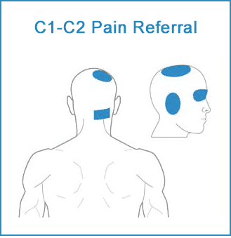
The Axial Cervical Compression test is primarily used to assess for nerve root involvement in the cervical spine. While it’s not directly related to assessing jaw pain, it can provide valuable information in certain cases where cervical spine pathology may be contributing to or mimicking jaw pain. Involvement of the cervical facet joints can lead to localized neck pain or cause pain referral to the skull. As shown below, the C1-C2 facet joints may cause a pain referral to the preauricular, occipital, and orbital regions, as well as the vertex. The negative result of this patient’s Axial Cervical Compression test suggests that the upper cervical facet joints are not involved.
Please watch the video below to see how the Axial Cervical Compression test is performed.
Reflection Point
Please stop and take a moment to consider whether all the elements of an adequate and relevant physical examination have been completed for this patient. Are there any additional procedures you would have performed and, if so, why?
Diagnosis
Based on the available information, what is the most likely diagnosis?
View Suggested Answer
Right TMJ dysfunction.
References and Suggested Further Readings:
Pavia S. et al. Chiropractic Treatment of Temporomandibular Dysfunction: A Retrospective Case Series. J Chiropr Med. 2015 Dec;14(4):279-84.
Lomas J. et al. Temporomandibular dysfunction. AJGP. Volume 47, Issue 4, April 2018.
Travell J. Temporomandibular joint pain referred from muscles of the head and neck. J Pros Dent. 1960;10:745.
Shanavas M. et al. Transcutaneous electrical nerve stimulation therapy: An adjuvant pain controlling modality in TMD patients – A clinical study. Dent Res J (Isfahan). 2014 Nov;11(6):676-9.
Ba S. et al. Ultrasound is Effective to Treat Temporomandibular Joint Disorder. J Pain Res. 2021 Jun 10;14:1667-1673.
Thank you for Reviewing this Free Sample.
This unit also has a 10 question multiple-choice quiz. The quiz is not included in this free sample.
Earn CE or CPD hours
in the comfort of your own home
and at your own pace.


Disclaimer: The Educom Continuing Education™ website (including text, graphics, downloadable resources, and videos) is intended to provide general health information for educational purposes only. This information is not a substitute for personal consultation with a qualified healthcare professional. Always seek the advice of a healthcare professional for any questions regarding your condition, symptoms, and appropriate treatments.
Copyright © Educom Pty Ltd: All content on the acemsk.com website, including text, graphics, videos, and downloadable files, is the property of Educom Continuing Education, a division of Educom Pty Ltd, and is protected by copyright and other intellectual property laws under international conventions. Unauthorized use or duplication of this material without express written permission from Educom Pty Ltd is strictly prohibited.